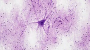
In the late 1800s, Spanish neuroscientist Santiago Ramón y Cajal drew hundreds of images of neurons. His exquisite work influenced our understanding of what they look like: Cells with a bulbous center, a forest of tree-like branches on one end, and a long, smooth tail on the other.
Centuries later, these images remain textbook. But a controversial study now suggests Ramón y Cajal, and neuroscientists since, might have missed a crucial detail.
A team from Johns Hopkins University found tiny “bubbles” dotted along the long tail—called the axon. Normally depicted as a mostly smooth, cylindrical cable, axons may instead look like “pearls on a string.”
Why care? Axons transmit electrical signals connecting the neural networks that give rise to our thoughts, memories, and emotions. Small changes in their shape could alter these signals and potentially the brain’s output—that is, our behavior.
“Understanding the structure of axons is important for understanding brain cell signaling,” Shigeki Watanabe at the Johns Hopkins University School of Medicine, who led the study, said in a press release.
The work took advantage of a type of microscopy that better preserves neuron structure. In three types of mouse neurons—some grown in petri dishes, others from adult mice and mouse embryos—the team consistently saw the nanopearls, suggesting they’re part of an axon’s normal shape.
“These findings challenge a century of understanding about axon structure,” said Watanabe.
The nanopearls weren’t static. Adding sugar to the neurons’ liquid environment or stripping neurons of cholesterol in their membranes—the fatty protective outer layer—altered the nanopearls’ size and distribution and the speed signals traveled down axons.
Reactions to the study were split. Some scientist welcomed the findings. Over the last 70 years, scientists have extensively studied axon shape and recognized its complex structure. With improving microscope technologies, discovering new structures isn’t surprising, but it is rather exciting.
Others are more skeptical. Speaking to Science, Christophe Leterrier of Aix-Marseille University, who was not involved in the study, said: “I think it’s true that [the axon is] not a perfect tube, but it’s not also just this kind of accordion that they show.”
Cable With a Chance of Stress Balls
Axons stretch inches in the brain with diameters 100 times thinner than a human hair. Although mostly tubular in shape, they’re dotted with occasional bubbles, called synaptic varicosities, that contain chemicals for the transmission of information with neighboring neurons. These long branches mainly come in two types: Some are wrapped in fatty sheaths and others are “bare,” without the cushioning.
Although often compared to tree branches, axons are shapeshifters. A brief burst of electrical signaling, for example, causes synaptic varicosities to temporarily expand by 20 percent. The axons also grow slightly wider for a longer period, before settling back to their normal size.
These tiny changes have large impacts on brain computation. Like an electrical cable that can change its properties, they fine-tune signal strength between networks, and in turn, the overall function of neurons.
Axons have another trick up their sleeves: They shrink up into “stress balls” with injury, such as an unsuspected blow to the head during sports, or in Alzheimer’s or Parkinson’s disease. Stress balls are relatively large compared to synaptic varicosities. But they’re transient. The structures eventually loosen and regain a tubular shape. Rather than harmful, they likely protect the brain by limiting damage to smaller regions and nurture axons during recovery.
But axons’ shape-shifting prowess is temporary and often only under duress. What do axons look like in a healthy brain?
Pearls on a String
Roughly a decade ago, Watanabe noticed tiny bubbles in the axons of roundworms while developing a new microscopy technique. Although the structures were much smaller and more tightly packed than stress balls, he banked the results as a curiosity but didn’t investigate further. Years later, the University of Bergen’s Pawel Burkhardt also noticed pearly axons in comb jellies, a tiny marine invertebrate.
In the new study, Watanabe and colleagues revisited the head-scratching findings, armed with a newer microscopy technique: High-pressure freezing. To image fine details in the brain, scientists usually dose it with multiple chemicals to set neurons in place. The treated brains are then sliced extremely thin, and the pieces are individually scanned with a microscope.
The procedure takes days. Without care, it can distort a neuron’s membrane and damage or even shred delicate axons. In contrast, high-pressure freezing better locks in the cell’s shape.
Using an electron microscope—which outlines a cell’s structure by shooting beams of electrons at it—the team studied “bare” axons from three sources: mouse neurons grown in a lab dish and those from thin slices of adult and embryonic mouse brains.
All axons had the peculiar pearl-like blobs along their entire length. Roughly 200 nanometers across, the nanopearls are far smaller than stress balls, and they’re spaced closer together. The beads likely form due to biophysics. Recent studies show that under tension, sections of a long tube crumple into beads—a phenomenon dubbed “membrane-driven instability.” Why this happens and its impact on brain function remains mostly mysterious, but the team has ideas.
Seeing Is Believing?
Using mathematical simulations, they modeled how changes in the surrounding environment impacts an axon’s pearling and its electrical transmission.
Axons are surrounded by a goopy, protective protein gel, like a bubble suit. But they still experience physical forces—like when we rapidly snap our heads. Simulations found that physical tension surrounding neurons is a key player in managing axon pearling.
In another test, the team stripped cholesterol from the neurons—a component in their membranes—to make them more flexible and fluid-like. The tweak lessened pearling in simulations and slowed electrical signals as they passed through the simulated axon.
Recording electrical signals from living mouse neurons led to similar results. Smaller and more compactly packed nanopearls slowed signals down, whereas axons with larger and widely spaced ones led to faster transmission.
The results suggest an “intriguing idea” that changing biophysical forces could directly alter the speed of the brain’s electrical signaling, wrote the authors.
Not everyone is convinced.
Some scientists think the nanopearls are an artifact stemming from the preparation process. “While quick freezing is an extremely rapid process, something may happen during the manipulation of the sample” to cause beading, Pietro De Camilli at the Yale School of Medicine, who was not involved in the study, told Science. Others question if—like a stress ball—the nanopearls form during stress and will eventually unfold. We don’t yet know: Microscopy is a snapshot in time, rather than a movie.
Despite pushback, the team is turning to human axons. Healthy human brain tissue is hard to come by. They plan to look for signs of nanopearls in brain tissue removed during epilepsy surgery and from those who passed away due to neurodegenerative diseases. Brain organoids, or “mini-brains” developed from healthy people could also help decipher axon shape.
Regardless, the study spurs the question: When it comes to brain anatomy, what else have we missed?
Image Credit: Bioscience Image Library by Fayette Reynolds on Unsplash
* This article was originally published at Singularity Hub

0 Comments